
Diagnostic Imaging
Diagnostic Imaging
Prince Frederick, MD 20678
Fax: 1.888.906.8773
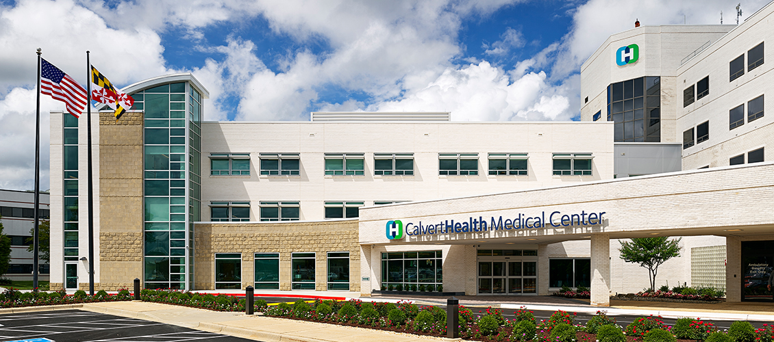
Through CalvertHealth’s partnership with American Radiology, diagnostic imaging services are offered through Calvert Medical Imaging Center. Conveniently located in the Calvert Medical Arts Building on the medical center campus, Calvert Medical Imaging Center brings you a broad range of services including MRI (3T Wide Open), MRA, CT, 3D mammography, 3D and 4D ultrasound, DEXA bone density scan, X-Ray, PET/CT imaging and more. Dedicated specialty physicians and cutting edge technology allow Calvert Medical Imaging Center to offer patients the very best in diagnosis, management, follow up and care.
Expand the sections below to learn more about the various imaging services related to the most prevalent cancer types in our region:
Learn More
Breast cancer is the second most common cancer in women (after lung cancer). Early detection of breast cancer vastly increases survival rates. Scheduling an annual mammogram after the age of 40 (or earlier, depending on your breast cancer risk factors) is one of the most important things women can do to ensure to take control of their health.
Some Breast Imaging Procedures Include:
Breast MRI
Some women — because of their family history, a genetic tendency or certain other factors — should be screened with MRI in addition to mammograms. The number of women who fall into this category is small: less than 2 percent of all the women in the United States. Talk with your doctor about your history and whether you should have additional tests.
Breast MRI helps to detect small lesions sometimes missed by mammography — without using radiation or compressing the breasts. MRI uses powerful magnets and radio waves to take very clear pictures of soft tissues, so it can be extremely useful in breast imaging.
Breast Ultrasound
Ultrasound uses sound waves instead of X-rays to exam breast tissue. A transducer is passed over the breast. The transmitted sound waves are translated into a picture on a monitor. It does not cause discomfort and, because it does not use radiation, it carries very little risk.
Ultrasound is useful for women with dense breasts or for evaluating suspicious areas seen by mammography or felt during a breast exam. It can also find breast lesions that are close to the chest, where mammography is less useful. Breast ultrasound can distinguish between cysts, which are fluid-filled, versus other types of solid breast masses.
Lower Dose 3D Mammography
3D mammography (tomosynthesis) is an FDA-approved technology that complements conventional 2D mammography. While traditional mammography generates 2D images, 3D mammography creates multiple thin 3D image slices that allow each section of the breast tissue to be seen more clearly.
New imaging software used with our 3D mammogram units enables us to offer a lower-dose 3D exam. The software eliminates the need to obtain additional 2D images as part of the 3D exam, as previously required. This reduces the radiation dose, making it similar to that of a traditional 2D mammogram.
A 3D mammogram looks and feels like a regular mammogram. An x-ray arm scans over your breast taking multiple 3D images at various angles. Each scan takes about 4 seconds and the entire 3D mammogram lasts about 15-20 minutes.
Stereotactic Breast Biopsy
Stereotactic biopsy uses mammography to precisely guide clinicians to a lump or abnormality such as an area of calcification, that cannot be felt or seen on ultrasound. Clinicians see a 3D picture of the lump’s exact location.
Image-guided biopsy allows patients to avoid hospitalization and general anesthesia (previously necessary with traditional surgical biopsy). It is a valuable method of getting a fast, accurate, conclusive diagnosis—without unnecessary time, pain or expense to the patient.
PET/CT
PET/CT combines Positron Emission Tomography (PET) with Computed Tomography (CT) technology to aid in the diagnosis of cancer, determining the extent to which cancer has spread, and assessing how a patient is responding to treatment. In addition, PET can be used to evaluate patients with cardiac and neurological disorders, such as seizures and dementia.
PET/CT is a nuclear medicine test that uses special imaging cameras and a radioactive solution called FDG (Flurodeoxyglucose) to create very clear pictures of a cell’s metabolism and if it is abnormal. CT uses X-rays to provide detailed anatomical information, including the location, size and shape of lesions or tumors in the body. When both applications are combined together, the two techniques provide accurate and specific information about where abnormalities are located and whether they are cancerous.
CT Scan
What is a CT scan?
A CT (computed tomography) scan is an advanced type of X-ray exam. Multiple X-rays are taken rapidly from a number of different angles around the body and then arranged by a high-speed computer to produce a cross-sectional view. CT may be used to visualize internal organs, head, neck, spine or extremities.
For some CT scans, the radiologist injects intravenous contrast medium or dye to highlight certain tissues for closer examination. Certain patients may also be required to drink oral contrast as well. A CT scan helps differentiate between healthy and diseased tissue, making it possible to accurately diagnose many diseases in their early stages.
When is a CT scan used?
CT scanning is generally used when your doctor needs more detailed diagnostic information than is possible from regular X-ray studies.
What happens during a CT scan procedure?
You will be positioned onto the table for the scan. You will feel the table move after each scan and may hear a whirring noise or high-pitched beep.
To get the most precise results, the technologist may ask you to hold your breath for a short time. Lie as still as possible to avoid blurring the images. You will be able to communicate with the technologist at all times during your scan. Actual time to acquire the images is often less than two minutes, but you should expect to be in the imaging room for approximately 10 to 15 minutes in its entirety.
You may leave immediately after your CT scan. If contrast was used, drink plenty of fluids, especially water, for the next 24 hours to help flush the contrast medium from your body. The radiologist will review your scans and send the results to your physician. Urgent findings will be called or faxed in to your physician very shortly after completion of your study.
What are the benefits and risks of a CT scan?
CT scans are among the safest exams we do. Your body will be exposed to a very small amount of radiation. If you are pregnant, you should not have a CT scan without first discussing the risks with your doctor. There is a small risk you will have an allergic reaction to contrast dye. Common minor allergic reactions may include hives and itching. More serious contrast reactions are very rare. Be sure to tell your health care provider if you know you are allergic to any medications or chemicals such as iodine. Our staff and physicians are trained and prepared to handle immediately any allergic reaction you might have and prevent any future occurrences.
Lower Dose Lung CT Screening
Lung cancer CT screening is one of the most accurate diagnostic tools for finding lung cancer at an early stage, when it is most treatable. CT scans of the lung are able to detect small abnormalities in the lungs that could be the beginning stages of lung cancer. These indicators are often not visible on a routine chest X-ray. Since a CT lung screening offers the best opportunity for successful treatment of lung cancer before symptoms are noticed, more physicians are opting for lung cancer screening based on risk factors (like smoking and family history), rather than symptoms.
PET/CT
PET/CT combines Positron Emission Tomography (PET) with Computed Tomography (CT) technology to aid in the diagnosis of cancer, determining the extent to which cancer has spread, and assessing how a patient is responding to treatment. In addition, PET can be used to evaluate patients with cardiac and neurological disorders, such as seizures and dementia.
PET/CT is a nuclear medicine test that uses special imaging cameras and a radioactive solution called FDG (Flurodeoxyglucose) to create very clear pictures of a cell’s metabolism and if it is abnormal. CT uses X-rays to provide detailed anatomical information, including the location, size and shape of lesions or tumors in the body. When both applications are combined together, the two techniques provide accurate and specific information about where abnormalities are located and whether they are cancerous.
Interventional Radiology
Interventional radiologist use imaging techniques such as X-rays, MRIs (magnetic resonance imaging) scans, fluoroscopy (an X-ray procedure that makes it possible to see internal organs in motion), CT (computed tomography) scans and ultrasounds.
Interventional radiologists perform a broad range of procedures such as treating tumours, taking organ biopsies or placing stents by inserting tiny instruments and thin plastic tubes (catheters) into the body via an artery or vein. The images are used to guide the catheters and instruments to the exact area where the procedure or treatment is to be performed. This reduces the need for traditional (open) or keyhole (laparoscopic) surgery as treatment can be given via a small plastic tube about the size of a straw.
PET/CT
PET/CT combines Positron Emission Tomography (PET) with Computed Tomography (CT) technology to aid in the diagnosis of cancer, determining the extent to which cancer has spread, and assessing how a patient is responding to treatment. In addition, PET can be used to evaluate patients with cardiac and neurological disorders, such as seizures and dementia.
PET/CT is a nuclear medicine test that uses special imaging cameras and a radioactive solution called FDG (Flurodeoxyglucose) to create very clear pictures of a cell’s metabolism and if it is abnormal. CT uses X-rays to provide detailed anatomical information, including the location, size and shape of lesions or tumors in the body. When both applications are combined together, the two techniques provide accurate and specific information about where abnormalities are located and whether they are cancerous.
Transvaginal Ultrasound
Transvaginal ultrasound imaging of the abdomen uses sound waves to produce pictures of the structures and organs in and around a woman's uterus. Transvaginal (TV) ultrasounds are performed very much like a gynecologic exam and involves the insertion of an ultrasound transducer into the vagina to study the lining of the uterus. These exams are frequently used to evaluate the reproductive and urinary systems. Ultrasound is safe, noninvasive and does not use ionizing radiation.
CT Scan
What is a CT scan?
A CT (computed tomography) scan is an advanced type of X-ray exam. Multiple X-rays are taken rapidly from a number of different angles around the body and then arranged by a high-speed computer to produce a cross-sectional view. CT may be used to visualize internal organs, head, neck, spine or extremities.
For some CT scans, the radiologist injects intravenous contrast medium or dye to highlight certain tissues for closer examination. Certain patients may also be required to drink oral contrast as well. A CT scan helps differentiate between healthy and diseased tissue, making it possible to accurately diagnose many diseases in their early stages.
When is a CT scan used?
CT scanning is generally used when your doctor needs more detailed diagnostic information than is possible from regular X-ray studies.
What happens during a CT scan procedure?
You will be positioned onto the table for the scan. You will feel the table move after each scan and may hear a whirring noise or high-pitched beep.
To get the most precise results, the technologist may ask you to hold your breath for a short time. Lie as still as possible to avoid blurring the images. You will be able to communicate with the technologist at all times during your scan. Actual time to acquire the images is often less than two minutes, but you should expect to be in the imaging room for approximately 10 to 15 minutes in its entirety.
You may leave immediately after your CT scan. If contrast was used, drink plenty of fluids, especially water, for the next 24 hours to help flush the contrast medium from your body. The radiologist will review your scans and send the results to your physician. Urgent findings will be called or faxed in to your physician very shortly after completion of your study.
What are the benefits and risks of a CT scan?
CT scans are among the safest exams we do. Your body will be exposed to a very small amount of radiation. If you are pregnant, you should not have a CT scan without first discussing the risks with your doctor. There is a small risk you will have an allergic reaction to contrast dye. Common minor allergic reactions may include hives and itching. More serious contrast reactions are very rare. Be sure to tell your health care provider if you know you are allergic to any medications or chemicals such as iodine. Our staff and physicians are trained and prepared to handle immediately any allergic reaction you might have and prevent any future occurrences.
PET/CT
PET/CT combines Positron Emission Tomography (PET) with Computed Tomography (CT) technology to aid in the diagnosis of cancer, determining the extent to which cancer has spread, and assessing how a patient is responding to treatment. In addition, PET can be used to evaluate patients with cardiac and neurological disorders, such as seizures and dementia.
PET/CT is a nuclear medicine test that uses special imaging cameras and a radioactive solution called FDG (Flurodeoxyglucose) to create very clear pictures of a cell’s metabolism and if it is abnormal. CT uses X-rays to provide detailed anatomical information, including the location, size and shape of lesions or tumors in the body. When both applications are combined together, the two techniques provide accurate and specific information about where abnormalities are located and whether they are cancerous.
Prostate MRI
Magnetic resonance imaging (MRI) of the prostate uses a powerful magnetic field, radio waves and a computer to produce detailed pictures of the structures within a man’s prostate gland. It is primarily used to evaluate the extent of prostate cancer and determine whether it has spread. It also may be used to help diagnose infection, benign prostatic hyperplasia (BPH) or congenital abnormalities.
Transrectal ultrasound (TRUS)
A transrectal ultrasound (TRUS) may also be called prostate sonogram or endorectal ultrasound. It is used to look at the prostate and tissues around it. An ultrasound transducer (also called a probe) sends sound waves through the wall of the rectum and into the prostate and surrounding tissue. A computer analyzes the wave patterns (called echoes) as they bounce off the organs and converts them into an image that doctors view on a video screen.
Diagnostic Radiography
Utilizes electromagnetic radiation through which still images of any body part can be produced on digital equipment and transmitted to the PACS monitors for viewing and interpretation. These x-rays can be performed anywhere in the facility through the use of portable machines.
Fluoroscopy
Shows dynamic images or images in motion by way of a monitor. This is used for monitoring the gastrointestinaI tract, blood vessels, genitourinary tract, needle guidance for injections, pacemaker insertions, orthopedic surgeries and much more. This same technology is widely used in the operating room and pain management centers. Fluoroscopic procedures normally require the use of contrast media which is instilled, ingested, or injected during the procedure.
CT Scanning
Uses x-rays, detectors, and computer equipment to process algorithms in the formation of images. CT can detect subtle differences in tissue densities that may indicate abnormal findings. Our modern, multi-slice CT scanners play a large role in the stroke services that we offer as an accredited Bronze Award Stroke Center. CT procedures such as Liver, Lung, or Kidney Biopsies, aspirations, and drainages are also done in our CT suite.
MRI
Uses very strong magnetic fields to align hydrogen nuclei within the body tissue. The nuclei give off signals as they return to their normal state and these signals are gathered by small coils placed at the area of interest to generate an image. With advances in technology, MRI gives the best tissue contrast possible and it has become a top choice of musculoskeletal and neuroradiology imaging.
Ultrasound
Uses high frequency sound waves to provide real time imaging of soft tissues in the body. Ultrasound uses no ionizing or exposing radiation so, generally, it is considered safer than radiography and CT. Therefore, it is largely used for obstetrical imaging as well as for imaging of abdominal organs, heart, thyroid, and reproductive organs. Vascular ultrasound applies the same physics and technology to concentrate on vascular structures such as the carotid arteries, aorta, and various veins throughout the body . Thoracentesis and paracentesis studies are performed in our ultrasound area.
Nuclear Medicine
Involves the use of radionuclides or radiopharmaceuticals which are swallowed, inhaled, or injected into the body and tagged to be taken up in specific organs. As the nuclides decay in the area, they emit radiation which is picked up by the nuclear camera and transformed into images through computer processing programs. This modality is part of the basis of PET CT which is also performed on our campus in the Calvert Medical Imaging Center located in the CMAC building. We also do nuclear cardiology studies to include nuclear cardiac stress tests done in conjunction with the patient's cardiologist. Our Nuclear Medicine unit offers dual technology that includes CT to include best practice imaging in cardiac, gastrointestinal, orthopedic studies, and infectious diseases.
Interventional Radiology
Is the area commonly known as special procedures. These studies are done by board certified radiologists and include angiography which is the study of blood vessels, kyphoplasty to treat chronic spine pain, nephrology studies such as nephrostomy tubes and kidney biopsies, dialysis catheter insertion, stent insertion to include Medicare-approved carotid stenting, thrombectomy procedures, UFE or uterine fibroid embolization, and other procedures such as Mediport and central line insertions.
Central Venous Access or PICC services
Are available which indicates the use of a peripherally inserted central catheter that is a long, slender, flexible catheter usually inserted into the upper arm and advanced until the tip reaches a large vein near the heart. Ultrasound in used to verify placement and the procedure is usually followed by a chest x-ray to assure correct final placement. These lines are normally used for long-term antiobiotic treatment, multiple access, nutrition and they are less invasive and used for longer periods than other methods.
Interventional Radiology Clinic services
Are by appointment only during which time the Interventional Radiologist will meet with patients and families to discuss history, known pathologies, diagnostic testing, and treatment options. Patients are referred to the IR Clinic by their primary or consulting physicians.
Diagnostic Cardiac Catheterization
Includes the visualization of the heart and associated blood vessels via a catheter that is inserted into the femoral artery. Upon insertion, contrast media is injected and images are seen on the monitor. The purpose of diagnostic cardiac catheterization is to collect information about the heart and the condition of the organ and blood vessels that supply blood to the area. Special hemodynamic monitoring is performed during the catheterization to record pertinent information about the condition of the patient's heart and coronary vessels. Cardiac catheterizations are performed by board certified cardiologists.